The visual cortex is connected with the cerebellum at every level. There are cortico-ponto-cerebellar pathways that form multiple retinotopic visual maps in the cerebellum, including a large representation of the central visual field in the cerebellar vermis. Many of these cerebellar visual areas overlap with areas related to eye movements. The cerebellum is connected with all of the major cortical areas related to eye movements. The figure shows functional activation of the cerebellum from the visual cortex (especially area V5), parietal lobe (area IPS), and the frontal eye fields.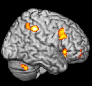 (figure from Kellermann et al 2012) The cerebellum gets involved in eye movements in a number of ways. The vestibulo-ocular reflex is probably the best studied, but each of the degrees of oculomotor freedom has a pathway through the cerebellum, and the cerebellum is vital for keeping the eyes on target. The cerebellum has three major pathways related to eye movements. The floccus and paraflocculus handle transient high frequency vestibular responses and get involved with smooth pursuit, gaze maintenance, and the VOR. They get direct input from interneurons in the abducens nucleus. The nodulus and ventral uvula process low frequency sustained vestibular input and are also involved with the VOR. The dorsal vermis and posterior fastigial nucleus contribute to the accuracy of saccades, in part by getting involved in the saccade termination process. The fastigial oculomotor area gets inputs from the burst and pause neurons in the reticular formation. 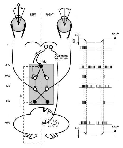 (figure from Robinson and Fuchs 2001) The main areas of the cerebellum involved in eye movements are the dorsal vermis and posterior fastigial nucleus, the nodulus and uvula, and the flocculus and paraflocculus (portions of the "medio-posterior cerebellum" including lobules VIc and VII). These areas connect with the oculomotor control system in the brainstem, including specifically the pause neurons in the nucleus prepositus raphe. The fastigial oculomotor region (FOR) in the caudal fastigial nucleus fine tunes the accuracy of horizontal saccades and contributes to smooth pursuit movements. The figure shows the overall anatomy and relationships to oculomotor areas.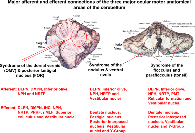 (figure from Shemesh and Zee 2019) Architecture of the CerebellumThe cerebellar circuits related to eye movements are organized like control systems. Since they integrate visual information with eye movements, they are naturally positioned in the area around T=0 on the timeline of brain electrical activity. The cerebellar circuits time eye movements on the basis of predictions related to the visual input. For example they match the eye velocity to the speed of moving visual targets. There are two main inputs to the cerebellum, the mossy fibers and the climbing fibers. The output from the cerebellum comes from the Purkinje cells. Inside the cerebellum is a very simple arrangement of neurons with behaviors that are mostly pretty well understood by now, although much is still being learned. The cerebellum has been best studied from the standpoint of motor control and coordination. Its architecture has been heavily conserved throughout evolution. The figure shows some of the major motor pathways through the cerebellum.  The figure below shows the characteristic internal architecture of the cerebellum. Mossy fibers enter from the precerebellar nuclei, and branch to connect with granule cells and Golgi cells in the specialized "dendritic claw" synapses, and with the deep cerebellar nuclei (both connections are excitatory). Each mossy fiber connects with about 50 granule cells. In turn, the deep cerebellar nuclei are also systemic outputs (they receive inhibitory inputs from the Purkinje cells) and project to premotor areas (mostly in the brainstem). The climbing fibers originate from the inferior olive, and branch to connect with the deep cerebellar nuclei and the dendrites of Purkinje cells. The cerebellar cortex therefore engages two other large structures, the deep cerebellar nuclei and the inferior olive. In the oculomotor system, the fastigial nucleus is one of the deep cerebellar nuclei. The fastigial oculomotor area is connected into both the targeting system in the superior colliculus, and the excitatory bursting neurons (EBNs) in the PPRF. Damage to the fastigial nucleus results in dissociable deficits, with dysmetric saccades and an offset in gaze direction during fixation. The gaze direction is related to the superior colliculus, whereas the saccade generating circuitry is in the PPRF (Guerrasio et al 2009).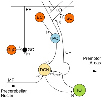 Much is still being learned about the cerebellum, even though it is one of the best studied areas in the brain. For example, it was previously thought that Golgi cells performed a smooth integration along the dendrites, but now it is clear they have voltage dependent calcium conductances and exhibit dendritic mini-spikes. Similarly it was once thought that Golgi cells were the only neurons that inhibit granule cells, but now it has been discovered that Purkinje cells directly inhibit granule cells in some specialized areas of the cerebellum, including those related to eye movements and vestibular function (Guo et al 2018). Golgi cells do not synapse directly onto Purkinje cells, that we know of - only onto granule cells via the dendritic claw synapses associated with the mossy fiber glomeruli.Next: Cerebral Cortex |